Explore the offer
Advanced Characterization and Fine Analysis / Electron, Ion and photon beam microscopy
Photo-Emitted Electron Microscopy
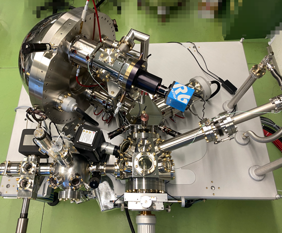
SPELEEM microscope uses low-energy electron as information carriers. Unlike scanning microscopy, electrons are collected simultaneously from an illuminated area of several tens of micrometers. The image formed by the magnification lenses can be acquired even in video-rate. SPELEEM is capable of combining microscopy, spectroscopy and diffraction to obtain a comprehensive characterization of conductive and semiconductive samples from the meso- to the nanoscale. Thanks to its versatility, SPELEEM can be employed to study model systems in the fields of nanotechnology, material science, catalysis, energy storage, organic and inorganic thin films, and low-dimensional materials.
SPELEEM is equipped with an hemispherical energy analyzer and two complemetary illumination sources - an electron gun and a He I/II UV lamp. Using the UV lamp, the following operational modes are available.
- Photo-Emission Electron Microscopy (PEEM): the photoemitted electrons are collected by the optical elements to form a magnified image on a 2D electron detector. Electrons can be filtered in energy and acceptance angle while traveling through the optical path. The contrast mechanism is given by the physics behind the photoemission process and the chemical state of the probed matter.
- Micron-sized Angle-Resolved PhotoEmission Spectroscopy (µARPES): by displaying the backfocal plane of the objective lens on the 2D electron detector, the microscope give access to a high-quality reciprocal space map of the photoemitted electrons. The probed kinetic energy of the photoemitted electrons can be filtered and varied to collect a complete characterization of the occupied band structure of the specimen. The photoemitted electron beam can be segmented to a few-micron sized area, enabling the collection of ARPES maps from a controlled portion of the sample.
Using an electron gun, LEEM mode can be implemented in the same instrument.
SPELEEM experimental facility can accommodate conductive and semiconductive samples hosted by Elmitec and Flag-style sample holders. Heating stages are provided in the microscope chamber and in the preparation chamber. Other equipment available includes a gas line with precision leak valves, e-beam evaporator, preparation chamber with noble-gas sputtering, fast entry lock for sample loading.
Available instruments
Select instruments to view their specifications and compare them (3 max)
Lab's Facility
Trieste